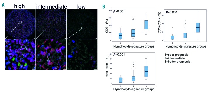
Figure 3.
Lower number of tumor-infiltrating T cells is associated with poor survival in primary testicular lymphoma (PTL). (A) Representative images (high, intermediate, low) from mIHC analysis of PTL tumor-associate macrophages probed with a 4-plex panel of T-cell markers. Blue: CD3; red: CD8; white: CD4; green: CD56; gray: DAPI. Scale bars 50 μm (upper panel) and 20 μm (lower panel). Images from individual channels are presented in Online Supplementary Figure S4. (B) Boxplots visualizing the expression of CD3+, CD3+CD4+, and CD3+CD8+ lymphocytes in the three groups based on the T-lymphocyte signature (1: poor prognosis, 2: intermediate, and 3: better prognosis). Statistical significance was determined by Kruskall-Wallis test.

