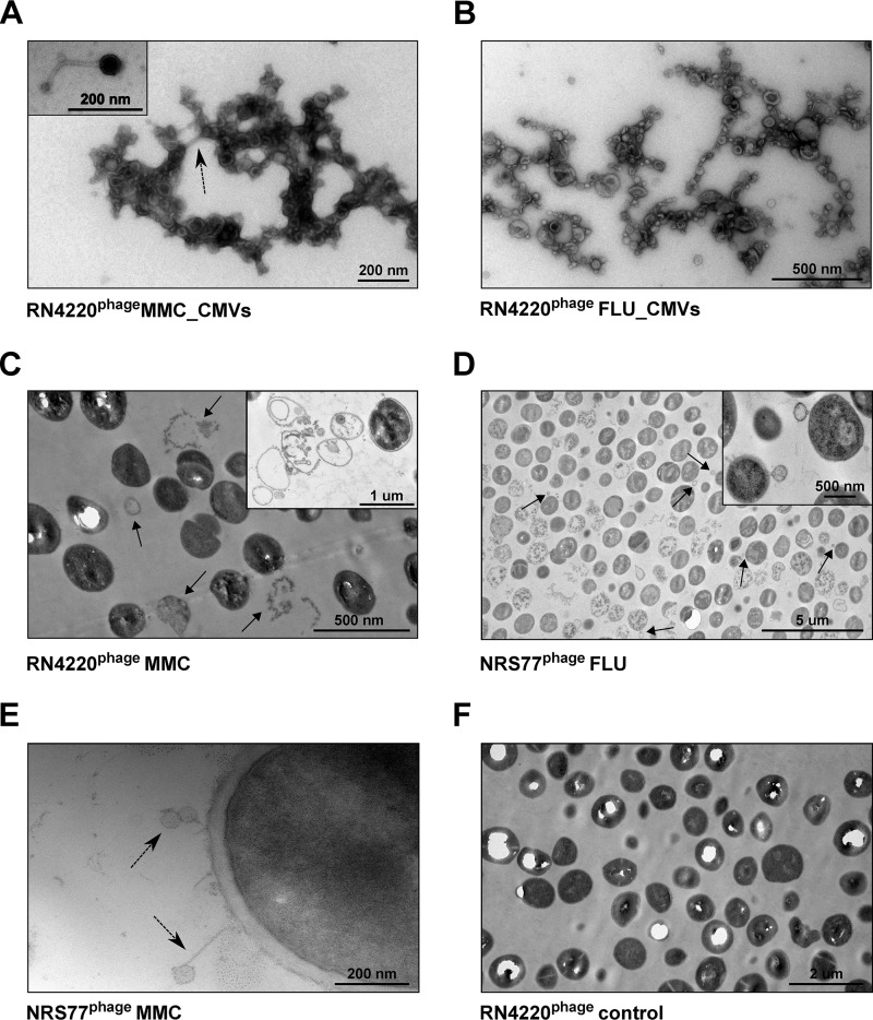
FIG 2.
TEM images of membrane vesicles and S. aureus after stimulation with MMC or FLU. (A through E) TEMs of CMVs and cells of S. aureus RN4220phage and NRS77phage treated with either 100 ng/ml MMC (A, C, E) or 10 times the MIC of FLU (B and D). Phages are indicated by dashed arrows. In samples of MMC-treated cultures, the presence of ghost cells (C) (indicated by arrows and shown in the inset) and of phages (E) was observed. In cultures treated with FLU, high numbers of ghost cells and cells with blebs (indicated by arrows and shown in the inset) were observed (D). (F) Untreated RN4220phage cells.

