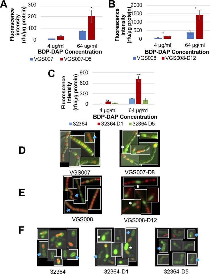
FIG 1.
BDP-DAP binding of S. mitis and S. oralis. Fluorescence intensity was normalized to protein ratio, using BDP-DAP binding to S. mitis VGS007 and VGS007-D8 (A), S. mitis VGS008 and VGS008-D12 (B), and S. oralis 32364, 32364-D1, and 32364-D5 (C). The increased binding of BDP-DAP is seen in DAP-R derivatives with mutations in cdsA compared to its DAP-S parental isolate. BDP-DAP staining (64 μg/ml) of Streptococcus cells is shown in green, propidium iodide (red), and overlay of DAP-S parental and DAP-R derivatives of VGS007 (D), VGS008 (E), and 32364 (F). Representative of uniform/septal binding is indicated by blue arrows, and hyperaccumulative binding is indicated by white arrows. *, P < 0.05; **, P < 0.01.

