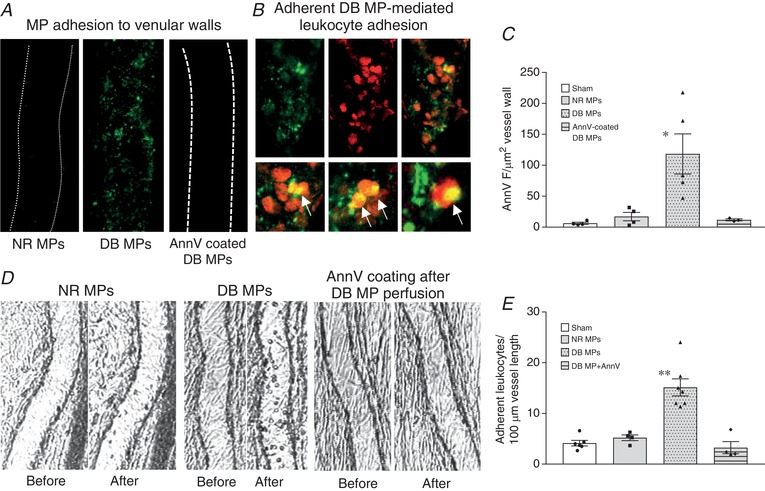
Figure 8. The externalized phosphatidylserine on diabetic microparticles (DB MPs) mediates their interactions with endothelium and circulating leukocytes.

A, representative confocal images show that only DB MPs (middle), not normal (NR) MPs (left), adhered to microvessel walls (marked green by AnnV labelling), even with matched MP concentrations. Blocking surface PS by pre‐coating DB MPs with AnnV prevented their adhesion (right). B, a representative vessel segment showing the adherent DB MP‐mediated leukocyte adhesion. Adherent MPs were marked by AnnV (green, top left) and adherent MP‐mediated leukocyte adhesion was labelled by Anti‐CD‐45 antibody (red, top middle). The top right image is the merged channel with local amplified regions shown in the lower row for details. C, summary of the quantification of AnnV fluorescence intensity (FI) at vessel walls as an indication of the number of adherent MPs of 4 groups (N = 5 for NR and DB MP group, N = 4 for AnnV pre‐coated DB MP group and sham control). D, isolated MP studies show that even with equal concentrations of MPs, only perfusion of diabetic MPs (N = 7), not normal MPs (N = 4), caused a significant leukocyte adhesion (left and middle images). Blocking the PS on adherent diabetic MPs on the vessel wall by perfusing AnnV into diabetic MP‐perfused vessels before resuming blood flow abolished diabetic MP‐mediated leukocyte adhesion (right images, N = 4). E, results summary for experiments presented in D (N = 6 for sham control). * P < 0.05 and ** P < 0.001 compared to the rest groups.
