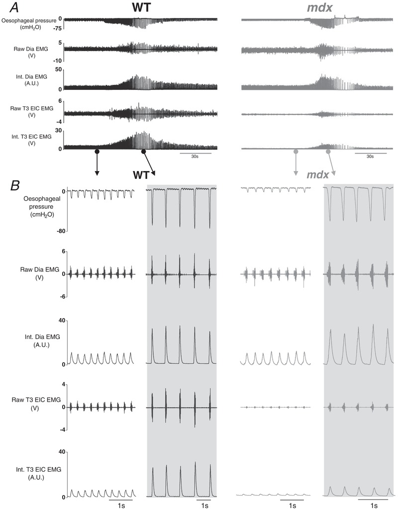
Figure 4. Oesophageal pressure and diaphragm and EIC EMG activities in anaesthetized wild‐type and mdx mice: original recordings.

A, representative recordings of oesophageal pressure and diaphragm (Dia) and T3 external intercostal (T3 EIC) raw and integrated (Int.) electromyogram (EMG) activity in a wild‐type (WT) (black) and mdx (grey) mouse during baseline ( = 0.60) and protracted tracheal occlusion (∼30–40 s). B, representative traces of oesophageal pressure and Dia and T3 EIC raw and Int. EMG activity in a WT and mdx mouse during baseline and peak inspiratory efforts (shaded) during a protracted tracheal occlusion.
