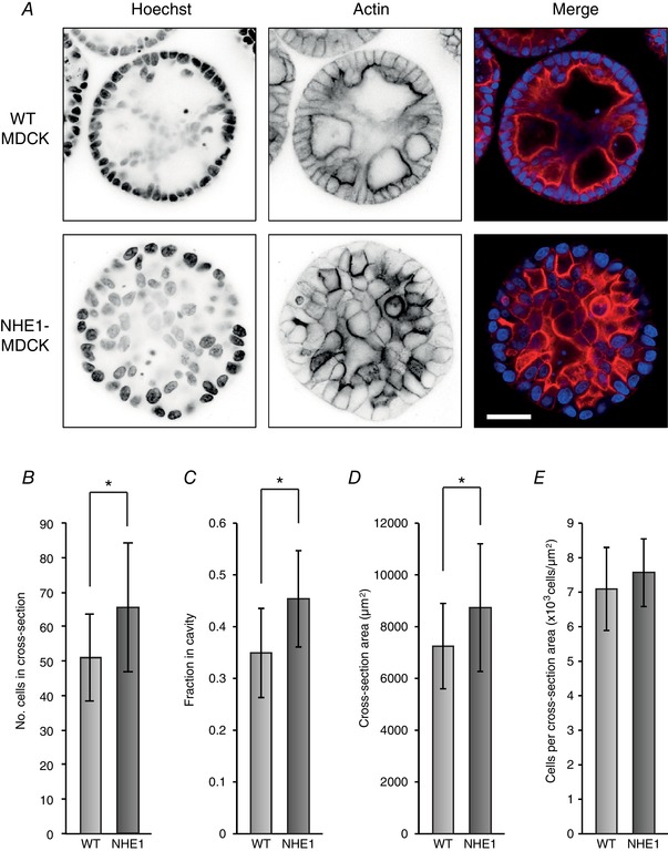
Figure 6. Polarity disruption in MDCK cysts overexpressing NHE1.

NHE1‐MDCK cells and WT MDCK cells were grown as cysts for 8 days. The cysts were then fixed and stained with Hoechst to visualize cell nuclei, as well as phalloidin to visualize actin. A, in cysts of WT MDCK cells, most cells were organized as a sphere with apical actin strands and cavities, although with some cells within the cyst. NHE1‐MDCK cysts were less organized with no or few cavities and no clear apical actin bands. B–E, cysts of NHE1‐MDCK cells contained more cells (B) and were larger (D), and more cells were localized in the cavity (C). However, there were similar numbers of cells relative to the size (E); n = 30 cysts (WT MDCK) and n = 28 cysts (NHE1‐MDCK) were included. * P < 0.05 using Student's t test. [Color figure can be viewed at wileyonlinelibrary.com]
