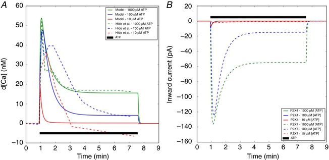
Figure 3. Predictions of Ca2+ increase and its corresponding inward currents via activation of P2X4 and P2X7 by ATP.

A, predicted free cytosolic Ca2+ (solid) with respect to time, subject to ATP treatments of 10–1000 μM ATP for 400 s (black), compared to experimental data from mouse microglia (dashed) (Hide et al. 2000) Complementary ER Ca2+ transients are reported in Fig. S5. B, inward current via P2X4 and P2X7 channels. Conditions to model the experimental conditions are listed in Table S2 (see also Sect. A.3.1 in Supporting Information). [Color figure can be viewed at wileyonlinelibrary.com]
