Figure 1.
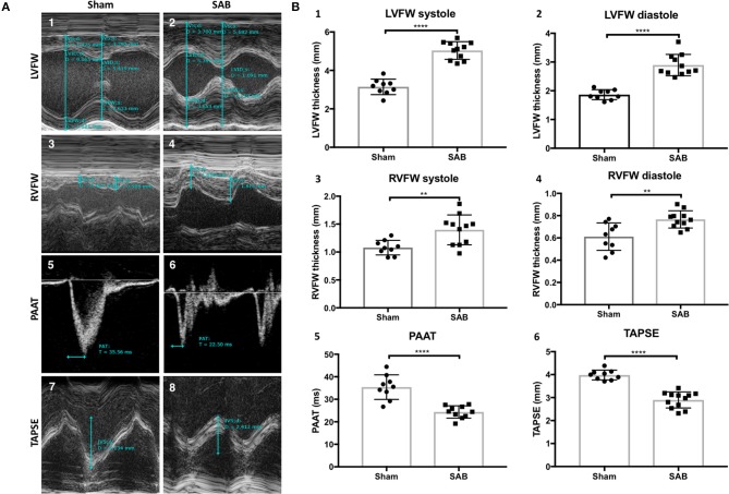
Echocardiographic measurements of sham vs. supra-coronary aortic banding (SAB) rats at 4 weeks post banding. (A) Representative echocardiographic images showing development of group 2 PH in SAB rats. (1–2) Left ventricular free wall thickness (LVFW), (3–4) right ventricular free wall thickness (RVFW), and (5–6) pulmonary artery acceleration time (PAAT), and (7–8) tricuspid annular plane systolic excursion (TAPSE) of sham vs. supra-coronary aortic banding (SAB) rats. (B) Summary statistics showing significant increase of biventricular thickness and decrease of PAAT and TAPSE in SAB vs. sham. (1) LVFW thickness, (2) diastolic LVFW thickness, (3) RVFW thickness, (4) diastolic RVFW thickness, (5) PAAT, (6) TAPSE of sham vs. supra-coronary aortic banding rats.

