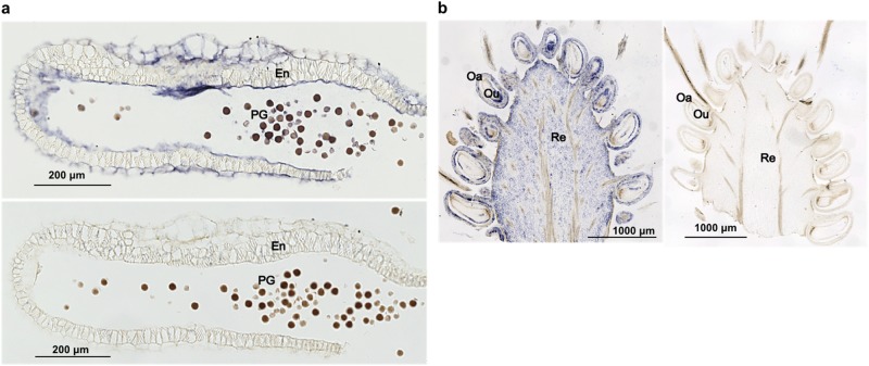
Fig. 4. In situ hybridization analysis of ALSV-AtFT distribution.
a ALSV distribution in the anther. ALSV was detected using an ALSV-Vp24(−) probe (top) and as a control experiment in the anther by using an SMV-P1(−) probe (bottom). Blue colour indicates distribution of ALSV in tissues. b ALSV distribution in fruit on plants infected with ALSV-AtFT detected by an ALSV-Vp24(−) probe (left). Control experiment in fruit using an SMV-P1(−) probe (right). PG pollen grain, En endothecium, Re receptacle, Oa ovary, Ou ovule

