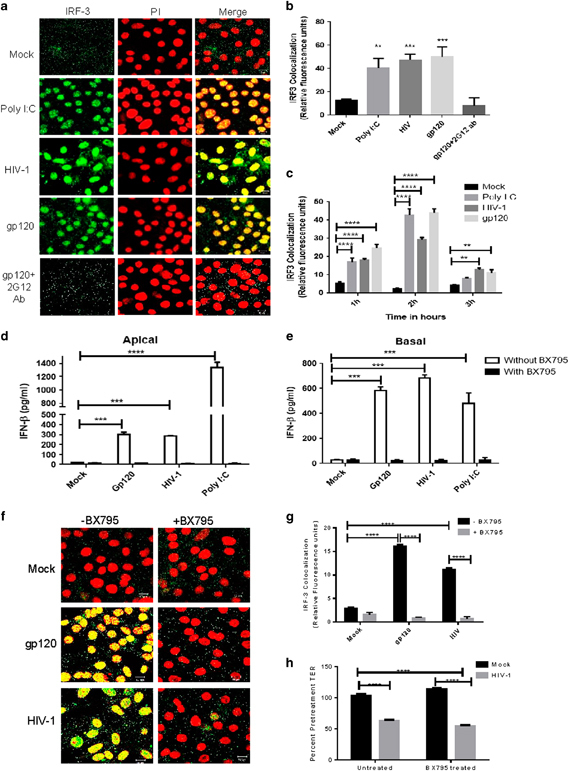
Figure 3.

Induction of IFNβ in endometrial GECs by HIV-1 gp120 is mediated through IRF3. Endometrial GECs were exposed to medium or poly I:C, HIV-1 (105 IU/well) or gp120 (100 ng/ml alone or with anti-gp120 neutralizing antibody) for 1–3 h. Cells were fixed and stained for the IRF3 (green fluorescence). Nuclear counterstaining (red fluorescence) was achieved using PI. Images were captured by a laser-scanning confocal microscopy. (a) Representative images are shown at 2 h time point from one of three separate experiments. Magnification × 1260. (b) IRF3 translocation and nuclear colocalization was measured by Image J software and presented as relative light units. (c) Time kinetics of IRF3 colocalization following treatment of endometrial GECs with medium or poly I:C (positive control), HIV-1 or gp120. (d, e) Endometrial GECs were incubated with the IRF3 inhibitor, BX795 (1 μM) for 1 h, before exposure with gp120, HIV-1 or poly I:C (positive control). Supernatants were collected after 24 h and assayed by ELISA. Results showed IFNβ production in apical (d) and basolateral supernatants (e). (f) Endometrial GECs were treated with BX795 for 1 h before gp120 or HIV-1 exposure for 2 h. The cells were fixed and stained for IRF3 and nuclei. Images were captured by laser-scanning confocal microscopy. Magnification: × 1260. (g) Colocalization was measured by image J software and represented in a bar diagram. h Endometrial GECs were preincubated with BX795 or media (mock) for 1 h and TERs were measured pretreatment and after 24 h of treatment with mock or HIV-1 to check whether BX795 was affecting HIV-1-mediated barrier disruption. Images are representatives of three separate experiments from cells isolated from three individual tissues. *P<0.05, **P<0.01, ***P<0.001 and ****P<0.0001. ELISA, enzyme-linked immunosorbent assay; GEC, genital epithelial cell; IFNβ, interferon-β; IRF3, interferon regulatory factor 3; PI, propidium iodide; poly I:C, polyinosinic:polycytidylic acid; TER, transepithelial resistance.
