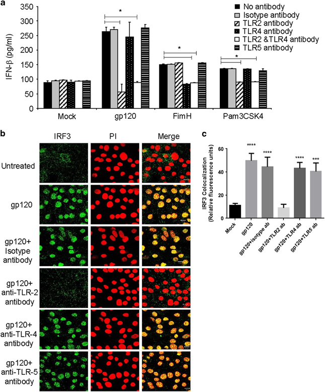
Figure 4.

Neutralization of TLR2 blocks gp120-mediated IFNβ production and IRF3 activation. (a) Endometrial GECs were pretreated with neutralizing antibodies against TLR2, TLR4, TLR5 or isotype control antibodies (all at 10 μg/ml) before exposure to gp120 (100 ng/ml) or mock treatment (media). FimH and Pam3CSK4 were used as positive controls for activation of TLR4 and TLR2, respectively. Supernatants were collected after 24 h and analyzed by ELISA for IFNβ production. Data shown are mean+s.d. and representative of three separate experiments done on cells isolated from three different tissues. (b) Epithelial monolayers were fixed after 2 h of exposure of gp120 with and without pretreatment with neutralizing antibodies against TLR2, TLR4, TLR5 or isotype control antibody and stained for IRF3. Propidium iodide was used to stain nuclei. Images were captures by a laser-scanning confocal microscopy. Magnification × 1260. Images are representative of one of three separate experiments done on cells isolated from three different tissues. (c) Quantitation of IRF3 colocalization were done by Image J software and presented in the graph. Significance was calculated by one-way ANOVA and IRF3 colocalization in all treatments were compared with mock treatment. *P<0.05, ***P<0.001 and ****P<0.0001. ANOVA, analysis of variance; ELISA, enzyme-linked immunosorbent assay; GEC, genital epithelial cell; IFNβ, interferon-β; IRF3, interferon regulatory factor 3; TLR, Toll-like receptor.
