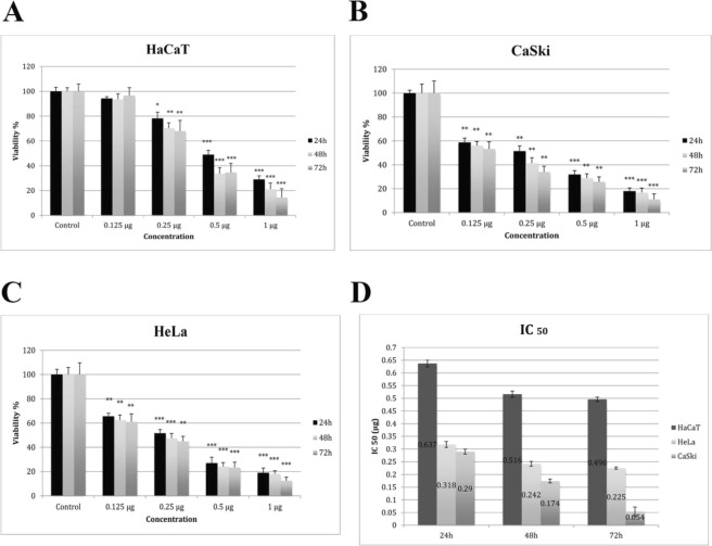
Figure 3.
0.125 µg Fig Latex showed cytotoxicity effects on CaSki (B), HeLa (C) but not on HaCaT (A) cells. Cell viability was assessed using MTT assay after 24, 48 and 72 hours of treatment with fig latex. Results represent the means of at least 3 independent experiments and were normalised to control wells. Error bars indicate SEM; *p < 0.05, **p < 0.01, ***p < 0.001. (D) Determination of IC50 values following treatment with fig latex. Results represent the mean of at least 3.

