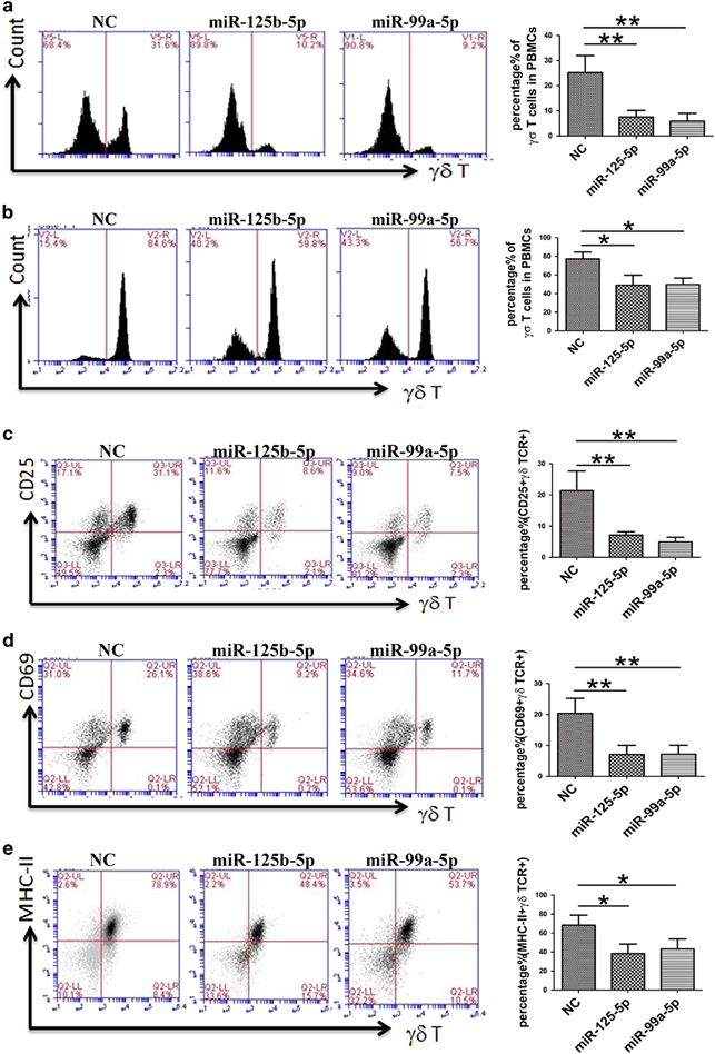
Figure 7.

miR-125b-5p and miR-99a-5p overexpression inhibited γδ T-cell activation. PBMCs were isolated from peripheral blood and infected by lentivirus expressing miR-125b-5p and miR-99a-5p. On days 7 (a) and 9 (b) after being stimulated with an anti-pan γδ TCR antibody in complete medium with IL-2, γδ T-cell activation was suppressed. The percentage of γδ TCR+ cells was greatly reduced. Even after 14 days, the γδ T-cell ratio did not exceed 70%. Simultaneously, expression of the activation markers was downregulated. Flow cytometry (FCM) was used to examine the expression levels of CD25 (c) and CD69 (d) on day 5 and of MHC-II (e) on day 9. The image in the right column shows the quantitative results. Data are shown as the means±s.d. (n=4 independent experiments). The differences (paired Student’s t-test) were significant (**P<0.01).
