
Figure 5.
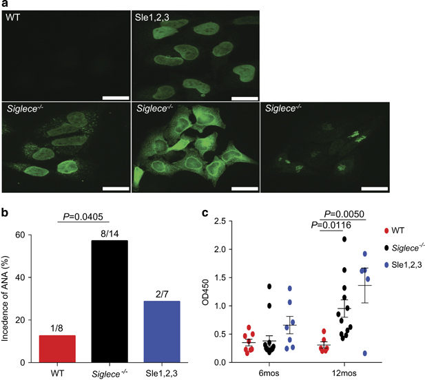
Increased production of autoantibodies in Siglec-E-deficient mice. (a) Autoantibody staining patterns using indirect fluorescence autoantibody assays in HeLa cells with sera derived from 6-month-old female WT, Siglece −/− and B6.NZM Sle1/Sle2/Sle3 (Sle1–3) mice at 1:1000 dilutions (scale bars, 20 μm; original magnification, x60). (b) Sera from 6-month-old female mice showed that ANA levels were markedly increased in Siglece −/− mice. WT, N=8; Siglece −/−, N=14; Sle1–3, N=7. (c) dsDNA autoantibody levels were assessed by ELISA with sera derived from 6-month-old and 12-month-old female mice. All sera were tested in the same assays. Each point represents a value from an individual mouse, and horizontal bars denote means with standard error of the mean (s.e.m.). Sample sizes were as follows: 6 months: WT, N=8; Siglece −/−, N=14; Sle1–3, N=7. 12 months: WT, N=6; Siglece −/−, N=12; Sle1–3, N=5. P-values at 6 months were analyzed using Mann–Whitney tests and were found to be nonsignificant. P-values at 12 months were calculated with a two-tailed unpaired Student’s t-test. Errors bar show deviations in biological repeats. ANA, antinuclear antibody; dsDNA, double-stranded DNA; ELISA, enzyme-linked immunosorbent assay; Siglec, sialic acid-binding immunoglobulin-like lectin; WT, wild type.