Figure 6.

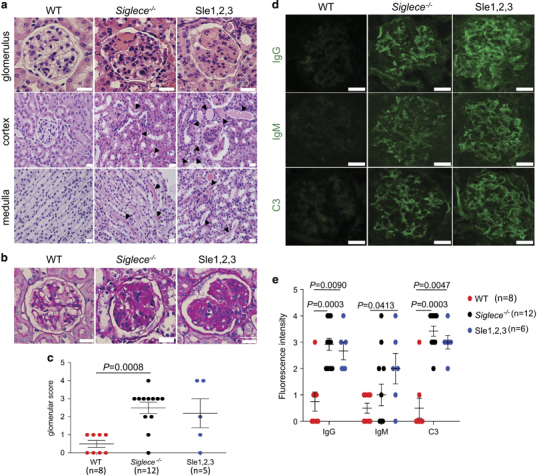
Increased renal pathology in Siglece −/− mice. (a) Kidney sections from 12-month-old female WT, Siglece −/− and B6.NZM Sle1/Sle2/Sle3 (Sle1–3) were stained with H&E. Arrows in Siglece −/− and B6.NZM Sle1/Sle2/Sle3 (Sle1–3) cortex and medulla indicate amyloid deposition. (b) PAS-positive material in the glomerular capillary lumen is shown in Siglece −/− and B6.NZM Sle1/Sle2/Sle3 (Sle1–3) mice. (c) Glomerular disease was scored from 0 to 4 for each mouse. Mice aged between 10–12 months were scored based on PAS staining. WT, N=8; Siglece −/−, N=12; Sle1–3, N=5. (d) Glomerular immune deposits were detected by immunofluorescence staining for IgG, IgM and complement C3. Representative images are shown (scale bars, 20 μm; original magnification, x60). Immunofluorescence staining was analyzed in triplicate. (e) Glomerular fluorescence intensity was scored from 0 to 4, in a double-blind manner. Each point represents a value from an individual mouse, and horizontal bars denote means with standard error of the mean (s.e.m.). WT, N=8; Siglece −/−, N=12; Sle1–3, N=6. P-values in (c and e) were calculated with a two-tailed unpaired Student’s t-test. Error bars show deviations in biological repeats. H&E, hematoxylin and eosin; Ig, immunogobulin; PAS, periodic acid-Schiff; WT, wild type.
