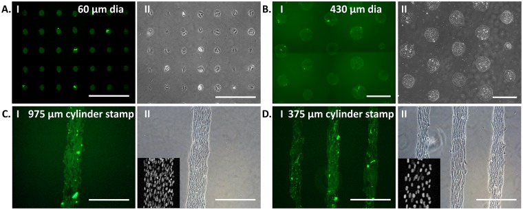
Figure 1.
Patterning of C2C12 myoblasts on FITC-conjugated collagen-I islands: Protein was printed using a stamp of 60 µm diameter fabricated with soft lithography (A-I) and manually fabricated in the lab, i.e., 430 µm diameter polystyrene beads stamp (B-I), 975 µm (C-I) and 375 µm (D-I) diameter PDMS cylinders stamp. C2C12, mouse myoblast, were then cultured on the protein patterned substrates using 60 µm diameter PDMS stamp (A-I I), 430 µm diameter polystyrene beads stamp (B-I I), 975 µm (C-I I) and 375 µm (D-I I) diameter PDMS cylinders stamp. On cylindrical patterns, the cells were aligned longitudinally. The nucleus of aligned cells stained with Hoechst is shown in the inset images (C-I I and D-I I). [Scale bar = 400 µm].

