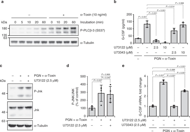
Fig. 3.
Activation of endogenous phospholipase C (PLC) by α-toxin contributes to increased production of granulocyte colony-stimulating factor (G-CSF). a Human umbilical vein endothelial cells (HUVECs) were cultured in the presence or absence of α-toxin, and whole-cell extracts were analyzed at the indicated time by immunoblotting with specific antibodies. Representative blots are shown of three independent experiments, raw gel images are available in Supplementary Figure 9. b–e HUVECs were cultured for 24 h (b), 1 h (c, d), or 4 h (e) in the presence of 10 ng ml−1 α-toxin and 10 μg ml−1 peptidoglycan (PGN) and in the presence or absence of the indicated concentration of U73122 or U73343. G-CSF levels in the culture medium were determined (b, n = 3 per condition). Whole-cell extracts were analyzed by immunoblotting with specific antibodies and the density of bands was measured (c, d, n = 4 per condition). Total RNA was extracted and subjected to real-time reverse transcriptase PCR using a specific primer set for G-CSF (e, n = 4 per condition). Representative blots are shown of four independent experiments, raw gel images are available in Supplementary Figure 9 (c). One-way analysis of variance was employed to assess statistical significance. Values are mean ± standard deviation. Similar results were obtained in two independent experiments

