Figure 6.

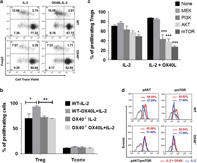
OX40 co-stimulation increases AKT-mTOR signaling to drive tTreg proliferation. (a) WT (top) and OX40−/− (bottom) CD8-CD4+SP thymocytes were treated with (right) or without (left) OX40L-Fc in the presence of IL-2 for 5 days. Numbers in the representative dot plots indicate percentages of resting (top right) vs proliferating (top left) CD25+Foxp3+ Tregs and resting (bottom right) vs proliferating (bottom left) CD25−Foxp3− Tconv cells. (b) Bar graph showing the percentages of proliferating Tregs and Tconv cells (values represent means±SEM, n=6, *p<0.05, **p<0.01). (c) Thymic CD4 SP cells were pre-treated with the indicated pharmacological kinase inhibitors (10 μM/ml) for 2 h and co-treated with IL-2 or OX40L+IL-2 for 5 days without any antigenic stimulation. Bar graph showing the percentages of proliferating Tregs in each of the culture conditions (values represent means±SEM, n=3, *p<0.05, ***p<0.005 vs No inhibitor control. (d) Histogram overlays showing MFI values of phospho-AKT and phospho-mTOR in Tregs expanded using IL-2 alone (blue) or IL-2+OX40L (red) in WT (top) vs OX40−/− (bottom) thymic CD4+ SP cells. Gray lines represent appropriate isotype controls. Treg, regulatory T cells
