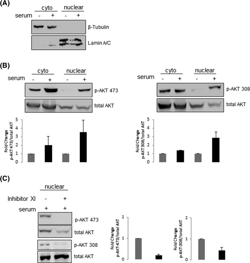
Figure 2. Both phosphorylated and non-phosphorylated AKT are present in melanoma cells nuclei.
A2058 cells were serum starved for 24 h and then stimulated (+) or not (−) with 10% serum for 30 min. (A) Nuclear and cytoplasmic fractions were analyzed by Western blot with anti-lamin A/C or anti-β-tubulin to confirm the purity of the nuclear and cytoplasmic fractions, respectively. (B) Nuclear and cytoplasmic fractions were analyzed by Western blot with anti-p-AKT-Ser473, anti-p-AKT-Thr308 and anti-total-AKT. (C) A2058 cells were serum starved for 24 h in the presence (+) or absence (−) of the PI3K inhibitor XI (20 μM) and then stimulated with 10% serum for 30 min (+). Nuclear fractions were analyzed by Western blot with anti-p-AKT-Ser473, anti-p-AKT-Thr308 and anti-total-AKT. (B and C) Results were plotted as the mean +/− S.D., n=3. Fold change in AKT phosphorylation was normalized to the levels of total-AKT.

