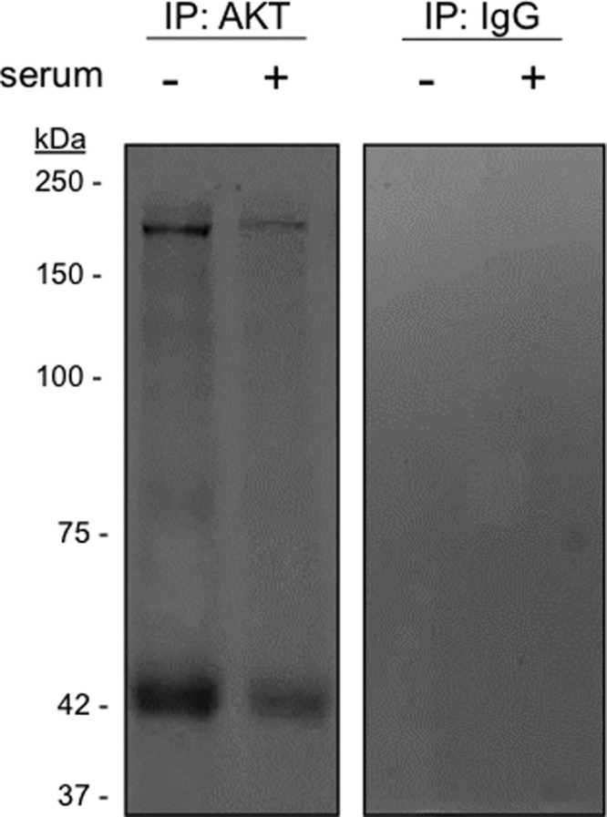
Figure 3. Identification of nuclear AKT binding proteins in melanoma cells nuclei.

A2058 cells were serum starved for 24 h and then stimulated (+) or not (−) with 10% serum for 30 min as indicated. Nuclear extracts were submitted to two-step chemical cross-linking and immunoprecipitations (IP) were performed using anti-total AKT or control IgG antibodies. Co-immunoprecipitated materials were resolved on SDS-PAGE. Coomassie blue staining of the gel revealed co-immunoprecipitated bands of approximately 42 and 200 kDa. The identified bands were excised and subjected to trypsin digestion for LC-MS/MS analysis.
