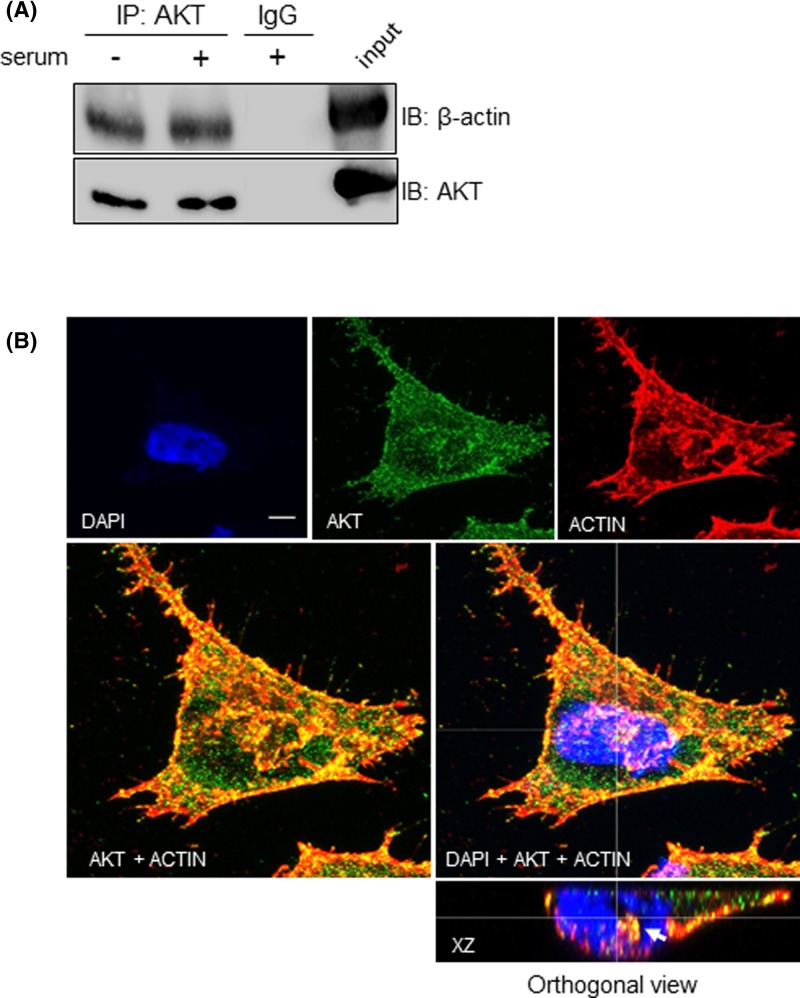
Figure 4. Nuclear AKT interacts with β-actin.
A2058 cells were serum starved for 24 h and then stimulated (+) or not (−) with 10% serum for 30 min, as indicated. (A) Nuclear lysates were submitted to immunoprecipitation (IP) with anti-total AKT or control IgG antibodies. Cell lysates prior to immunoprecipitation were used as input. Input and co-immunoprecipitated proteins were immunoblotted (IB) for detection of total AKT and β-actin. (B) A2058 cells were double immunostained for AKT and β-actin and analyzed by confocal laser-scanning microscopy. Confocal images show maximum projection of image stacks. AKT is shown in green, β-actin is shown in red, DAPI-stained nuclei are shown in blue. Merged images are shown at higher magnification in the panels below. Reconstructed orthogonal projection is presented as viewed in the xz plane, showing co-localization of AKT with β-actin in the nucleus (yellow pixels, arrowhead); scale bar, 5 μm.

