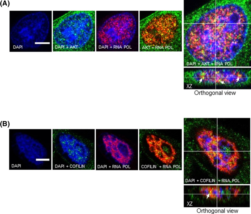
Figure 7. Both nuclear AKT and cofilin co-localize with active RNA Pol II.
A2058 cells were double immunostained either for (A) AKT or (B) Cofilin with RNA Pol II phosphorylated at Ser2 in the CTD. AKT and Cofilin are shown in green, RNA Pol II is shown in red and DAPI-stained nuclei are shown in blue. Immunostained cells were analyzed by confocal laser-scanning microscopy. Images are projections of one stack from the middle plane of the nucleus. Reconstructed orthogonal projections are presented as viewed in the xz plane, showing co-localized immunostaining in the nucleus (yellow pixels, arrowhead); scale bar, 5 μm.

