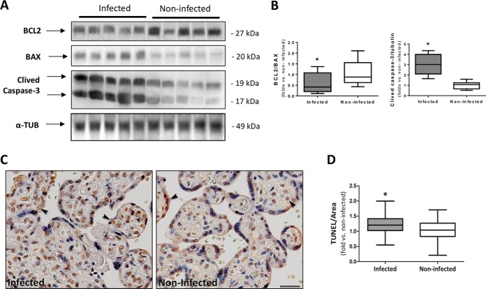
Fig. 7.
Higher apoptosis induction in past P. falciparum-infected placentas. A, Western blot analysis of BCL2, BAX, and Caspase-3 levels in past infected and noninfected placentas. α-tubulin was used as loading control and for normalization. The same cohort of sample from the mass spectrometry analysis was used for this assay. B, The BCL2/Bax and Caspase-3/Tubulin ratios were calculated by densitometry analysis (n = 5 infected and 5 noninfected, Student t test *p < 0.05). C, Apoptotic nuclei (TUNEL stain brown) in past-infected and noninfected placentas. Arrowheads show positive nuclei examples. The images in each panel were acquired at 400× magnification and the scale bar represents 25 μm. Mayer's hematoxylin counterstaining. Independent cohort of samples was used for this assay. D, Quantification of TUNEL immunostaining intensity (pixels/μm2) per tissue area is represented by number of fold-increase to noninfected mean (n = 24 infected and 32 noninfected placentas, Mann Whitney test *p < 0.05).

