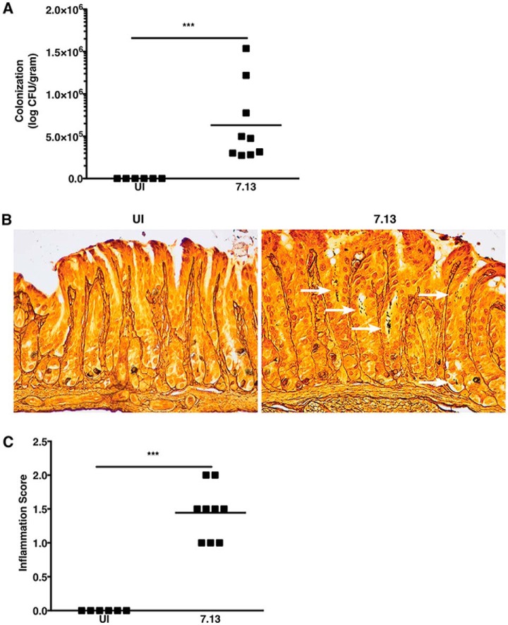
Fig. 1.
H. pylori strain 7.13 colonizes gerbils and induces inflammation. A, Gastric tissue from uninfected (UI) and H. pylori-infected gerbils was homogenized and plated on selective trypticase soy agar plates with 5% sheep blood for isolation of H. pylori. Plates were incubated for 3–5 days, and colonization density was determined and expressed as log colony-forming units (CFU) per gram of gastric tissue. Each data point represents colonization density from an individual animal. B, Linear strips of gastric tissue, extending from the squamocolumnar junction through the proximal duodenum, were fixed in 10% neutral-buffered formalin, embedded in paraffin, and stained with Steiner stain to identify H. pylori topography within gastric tissue sections. White arrows designate regions with H. pylori colonization. C, Linear strips of gastric tissue, extending from the squamocolumnar junction through the proximal duodenum, were fixed in 10% neutral-buffered formalin, embedded in paraffin, and stained with hematoxylin and eosin. A pathologist (MBP), blinded to the treatment groups, assessed indices of inflammation. Severity of acute and chronic inflammation was graded 0–3 (absent (0), mild (1), moderate (2), or marked (3) inflammation) in both the gastric antrum and corpus. Each data point represents inflammation scores from an individual animal. Mann-Whitney U test was used to determine statistical significance between uninfected and infected groups. ***, p < 0.005.

