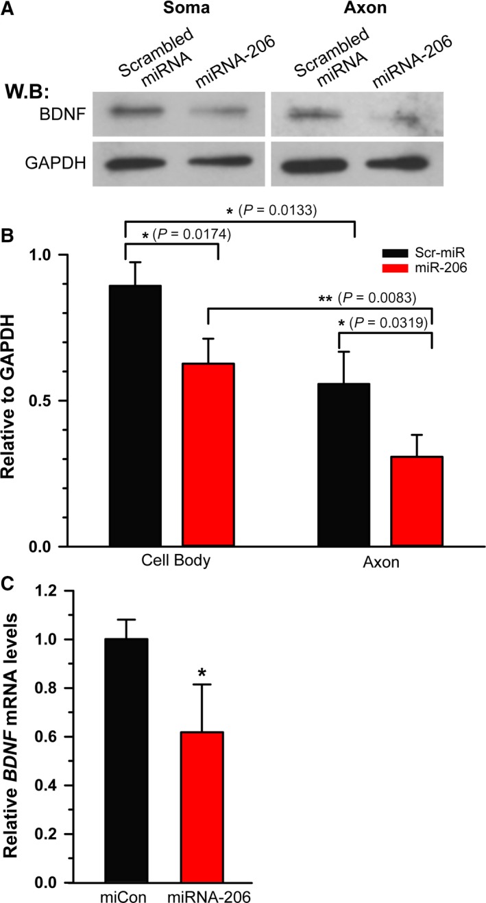
Figure 3.

miRNA‐206 decreases BDNF protein in sensory neurons. (A) Western blot analysis was performed on somal and axonal lysates obtained from transfected DRG neurons cultured for 3 days and probed with antibodies to BDNF and GAPDH. GAPDH was included as a loading control. (B) Densitometric analysis showed a significant decrease in both somal and axonal BDNF expression. Note that the decreased level of BDNF expression by miRNA‐206 in axon was statistically lower compared to that in soma. *P < 0.05, **P < 0.01 by Student's t‐test. Error bars represent the SD (n = 3). (C) Quantification of BDNF mRNA levels in miRNA‐206 transfected DRG neurons was measured by qRT‐PCR and normalized to GAPDH mRNA. Consistent with the reduction in BDNF protein, the significant reduction in BDNF mRNA level indicted miRNA‐206‐mediated regulation of BDNF expression at the mRNA level. *P < 0.05 by Student's t‐test. Error bars represent the SD (n = 3).
