
Figure 2.
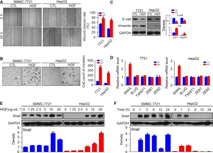
Hepatocyte growth factor induces EMT by up‐regulating Snail in HCC cells. (A) Serum‐starved SMMC‐7721 and HepG2 were stimulated with or without HGF (10 ng·mL−1) for 24 h, and then cell migration was determined by a wound healing assay. (B) Serum‐starved SMMC‐7721 and HepG2 were stimulated with or without HGF (10 ng·mL−1) for 24 h, and then cell invasion was determined by a transwell assay. (C) Serum‐starved SMMC‐7721 and HepG2 were stimulated with or without HGF (10 ng·mL−1) for 48 h, and the protein levels of E‐cadherin and vimentin were detected by western blotting. The density of each band was normalized to GAPDH. (D) Quantitative RT‐PCR results of snail, slug, twist1, zeb1 and zeb2 after incubation with HGF for 3 h. (E) Serum‐starved SMMC‐7721 and HepG2 were stimulated with HGF at different concentrations for 3 h, and protein levels of Snail were detected by western blotting. The density of each band was normalized to GAPDH. (F) Serum‐starved SMMC‐7721 and HepG2 were stimulated with HGF (10 ng·mL−1) for different times, and protein levels of Snail were detected by western blotting. The density of each band was normalized to GAPDH. (*P < 0.05, **P < 0.01, compared to control). Data are expressed as the mean ± SD from three individual experiments. Differences between groups were determined using Student's t‐test and two‐way ANOVA with Bonferroni correction.