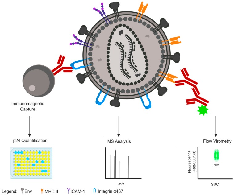
Figure 1.
Methods to detect host protein incorporation on the surface of an HIV-1 virion. An HIV-1 particle is depicted with surface antigens displayed, including the viral envelope glycoprotein (Env, grey) and host cell proteins MHC II (orange), CD54/ICAM-1 (purple), and integrin α4β7 (blue). Immunomagnetic bead capture (left) is one common and crude method to determine surface antigen expression on virion surfaces, quantified by p24 antigen ELISA readout. Mass spectrometry (centre) is another common and crude method to detect host protein incorporation that might offer enhanced sensitivity over immunocapture, but cannot distinguish the location of incorporated proteins from virion surface versus virion interior. Flow virometry (right) is an emerging technology for the flow cytometry-based detection of nanoparticles, permitting highly sensitive detection of unique virion populations that can be distinguished based on scattering properties (x-axis) and fluorescence expression (y-axis) within virions or via staining with fluorochrome-conjugated antibodies (maroon). This figure was created with BioRender.

