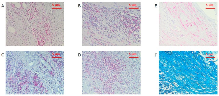
Figure 5.
Hot spots with tumor-infiltrating leukocytes of CD3+ (A), CD8+ (B), CD20+ (C), and CD66b+ (D) cells under a power field of 200× magnification as well as corresponding areas of α-sma (E) and collagen (F) stained spots. CD3 is the marker of T cells, CD8—of cytotoxic T cells, CD20—of B cells, CD66b—of neutrophils, and α-sma—of cancer associated fibroblasts.

