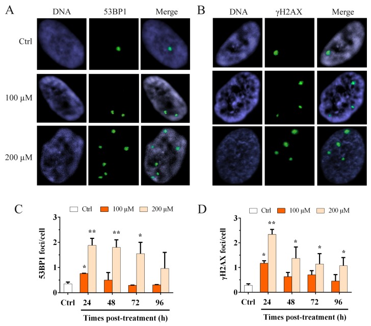
Figure 3.
Immunofluorescence staining for 53BP1 and γH2AX foci. Images of MRC-5 cells stained for (A) 53BP1 and (B) γH2AX. Immunofluorescence staining for 53BP1 and γH2AX was used to detect the activation of the genomic DDR. The columns show the frequency of (C) 53BP1 foci per cell and (D) γH2AX foci per cell, evaluated after 100 and 200 µM H2O2 treatment. At 24 h after treatment, we observed a significant increase in the 53BP1 and γH2AX foci for both doses. Conversely, for the higher dose, we showed a significant increase in both 53BP1 and γH2AX at 24 h that significantly persisted for up to 96 h. The bars denote the standard error. Statistical analysis was performed between treated and control samples. * p < 0.05; ** p < 0.01 by Student’s t-test.

