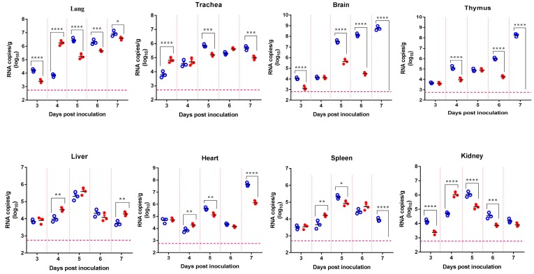
Figure 1.
Tissue distribution of Japanese encephalitis virus (JEV) RNA in mice infected by the intranasal route. Four-week-old mice were intranasally (i.n.) inoculated with 106.0 plaque-forming units (pfu)/50 μL of SCYA201201 or SA14-14-2 JEV virus. The viral RNA load was measured using real-time RT-PCR in each mouse tissue collected at 3, 4, 5, 6, and 7 days post-infection (dpi) and expressed as RNA copies per gram (Mean ± SEM) (○ represent the SCYA201201 strain, and ● represent the SA14-14-2 strain). The horizontal dotted lines indicate the limit of detection (* P ˂ 0.05, ** P ˂ 0.01, *** P ˂ 0.001, and **** P ˂ 0.0001).

