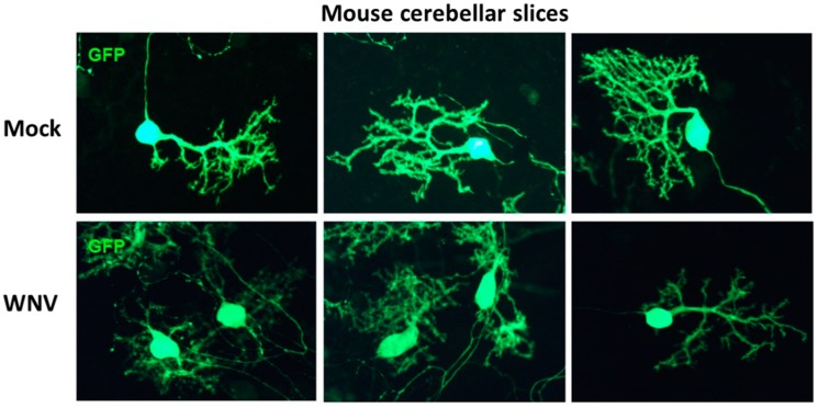
Figure 2.
GFP immunofluorescence in mock- or WNV-treated (107 PFU) cerebellar slice cultures. Since GFP localization, especially in the dendritic spines of Purkinje cells, is heterogeneous [26], the slices were immunostained with GFP antibody to enhance visualization. Purkinje cells (PCs) in WNV-exposed cultures show shrunken or pruned dendritic arbors and reduced GFP immunostaining as compared to mock cultures. The total GFP immunoreactive area of these images was quantified with ImageJ. The total area of WNV-treated PCs was significantly less (3736 ± 525.2, n = 5) than mock-treated PCs (9330 ± 1226, n = 3). The data are presented as mean ± SEM and p < 0.003.

