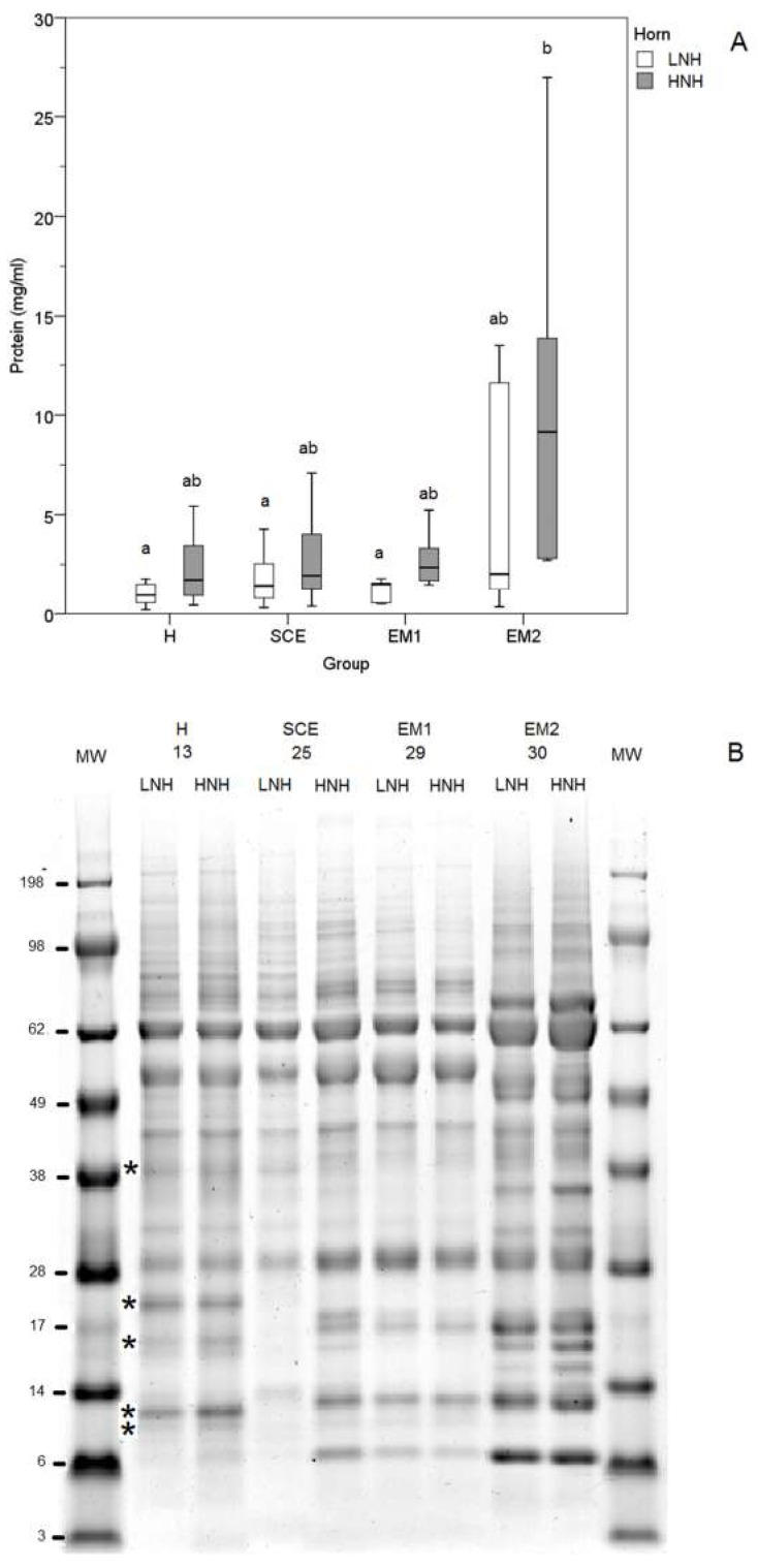
Figure 3.
Concentrations (A) and SDS-PAG electrophoresis (B) of the proteins in the uterine horn lavage (UHL) fluids obtained from the uterine horns with the higher and lower neutrophil percentage (HNH) and (LNH), respectively (A). MW: molecular weights; H (Healthy cows): Clinically healthy subjects with PMN < 5%, one week after the clinical evaluation; SCE (Subclinical Endometritis): Clinically healthy subjects and PMN ≥ 5%, one week after the clinical evaluation; EM1 (Grade 1 Endometritis): Animals with grade 1 clinical endometritis and PMN ≥ 5%, one week after the clinical evaluation; EM2 (Grade 2 Endometritis): Animals with grade 2 clinical endometritis and PMN ≥ 5%, one week after the clinical evaluation. Different superscripts (a, b) indicate a statistically significant different group (p < 0.05; Kruskal-Wallis one-way ANOVA and multiple comparisons, SPSS 24.0). The asterisks indicates electrophoretic bands exclusively present in the H cows.

