Figure 2.
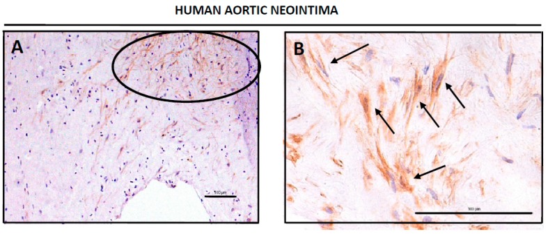
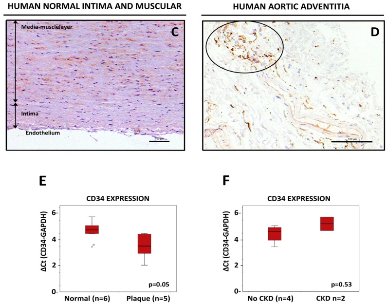
Detection of CD34-positive cells in human aortic tissue. Whole human arteries were isolated, sliced, prepared for IHQ analysis and stained for CD34 as described in Material and Methods. (A,B) Shown are representative images of two different neointimas, stained for anti-CD34, at different magnifications (20× in 3A, 40× in 3B). The circle in (A) shows a cluster of CD34-positive cells, and arrows in (B) show±± individual fibroblastoid cells positive for CD34 staining. Tissues were counterstained with hematoxilin-eosin. Bars are 100 μm. (C) Representative image of a human aortic tissue from a normal section (non-lesion) showing the intima and the muscular layer (vertical arrows) and stained for anti-CD34. Tissue was counterstained with hematoxilin-eosin. Bar is 100 μm. (D) Representative image of a human adventitia stained for anti-CD34. The circle in (D) shows a cluster of CD34-positive cells. Tissue was counterstained with hematoxilin-eosin. Bar is 100 μm. (E) Expression of CD34 mRNA measured by qPCR in human ATH plaques compared with normal abdominal aortas. (F) Expression of CD34 mRNA in aortic tissue with normal vascular walls from CKD patients when compared with non-CKD patients.


