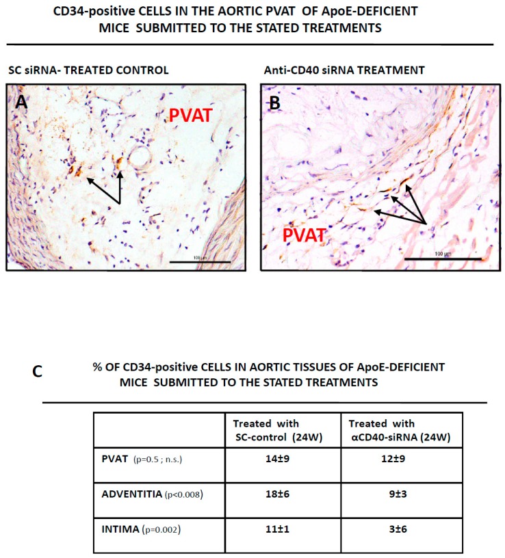
Figure 3.
Detection of CD34-positive cells in the murine aortic perivascular tissue. Aortas from ApoE-deficient mice treated with a scrambled (SC) siRNA for 24 weeks as control, or with an anti-CD40 specific siRNA also for 24 weeks, were isolated, sliced, prepared for IHQ analysis and stained for CD34 as described in Material and Methods. (A,B) Show representative fields with individual CD34-positive cells (arrows) in the SC-siRNA control (A) or in the anti-CD40 specific siRNA (B). Tissues were counterstained with hematoxilin-eosin. Bars are 100 μm. (C) Quantification of CD34-positive cells in the PVAT, intima and adventitia of aortas of ApoE-deficient mice submitted to the stated treatments. A total of 10 fields were examined by two different researchers and the result expressed as % of CD34-positive cells with regard of the total number of eosin-positive nuclei in the field.

