Figure 1.
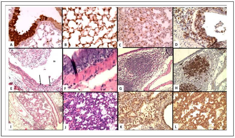
(A–C) Virus localization in epithelial cells. (D–F) T-Cell infiltrate in epithelial cells. (G,H) iBALT (I–L) Epithelial proliferation. (A) Immunoperoxidase staining of influenza virus in bronchial epithelium of WT mouse at 8 days post-infection (200×); (B) Virus in type II pneumocytes in alveoli of WT mouse (400×); (C) Decreased virus staining in type II pneumocytes with lymphocytic infiltrate in mice receiving memory T cells (400×); (D) CD3 T-cell staining of peri-bronchial T-cells and cells in bronchus of SCID mice 1 week after transfer of memory CD4 T-cells (400×); (E) H&E showing lymphocytes infiltration of bronchi and desquamated cells in WT mice 2 days after transfer of memory CD4 T-cells (100×); (F) H&E of intraepithelial lymphocytes in IL-10 knock-out (KO)mice, day 8 (400×); (G) H&E iBALT 5 days after secondary infection of Treg depleted mice (40×); (H) B-cell staining PAX5) of iBALT (100×); (I) H&E epithelial proliferation in SCID mice day 14 after transfer of memory CD4 T-cells (100×). (J–L) proliferating epithelial cells in Tregs depleted mice week 4; (J) H&E; K. surfactant; (L) TTF (200×).

