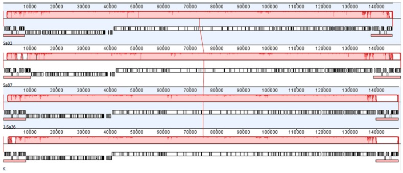
Figure 2.
A Progressive Mauve alignment of (top to bottom) Sa83, Sa87, J-Sa36, and phage K (GenBank K766114), each showing annotated genes (white boxes) and long terminal repeats (small red boxes immediately below white gene blocks). The large red blocks above each annotated genome (connected by the red vertical line at approximately 75 kb) represent local collinear blocks of genomes identity; interruptions in these red blocks indicate differences among the four aligned nucleotide sequences.

