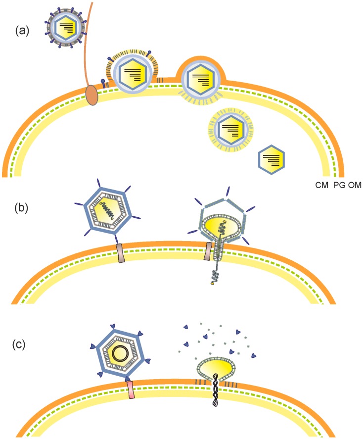
Figure 1.
Entry mechanisms of membrane-containing phages. (a) Pseudomonas phage phi6. Phi6 attaches to a type IV pilus, which retracts and brings the virion into contact with the bacterial outer membrane. The viral membrane (envelope) fuses with the host outer membrane, releasing the nucleocapsid into periplasmic space. The peptidoglycan layer is digested by a virally-encoded lytic enzyme, after which the nucleocapsid enters the cytoplasm via an endocytic-like route. Finally, the nucleocapsid shell dissociates, releasing the phi6 virion core (polymerase complex). (b) Pseudomonas phage PRD1. Upon attachment to the host receptor, the internal membrane vesicle of phage PRD1 transforms into a proteo-lipidic tube, which traverses the cell envelope and provides a conduit for transferring the linear dsDNA genome into the cytoplasm. (c) Pseudoalteromonas phage PM2. It has been suggested that after phage PM2 binds to the host receptor, its protein capsid dissociates triggering the fusion between the internal membrane vesicle and the bacterial outer membrane and consequently the release of the circular dsDNA genome into the cell. CM, cytoplasmic membrane; PG, peptidoglycan layer; OM, outer membrane of the envelope in Gram-negative host bacterium.

