Figure 3.
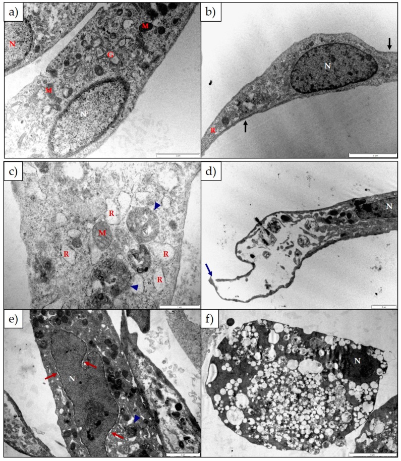
Electron micrograph of (a,b) untreated H9c2 cells and H9c2 cells exposed to (c) 25 µM and (d) 100 µM 1α,2α-epoxyscillirosidine for 24 h; (e) 100 µM 1α,2α-epoxyscillirosidine for 48 h; (f) 100 µM 1α,2α-epoxyscillirosidine for 72 h. The untreated cells were long and thin with tapered ends. The cytoskeleton was associated with the plasma membrane (black arrows). The H9c2 cells treated with 1α,2α-epoxyscillirosidine has plasma membrane damage (blue arrow), swollen RER and perinuclear spaces (red arrows) and cells were extensively vacuolated. Autophagic vesicles could be seen within the cytoplasm (blue arrow heads). G—Golgi Complexes; M—Mitochondria; N—Nuclei; R—Rough Endoplasmic Reticulum. The scale bar at the bottom right corner represents 5 µm (b,f), 2 µm (a,d,e) and 1 µm (c) respectively.

