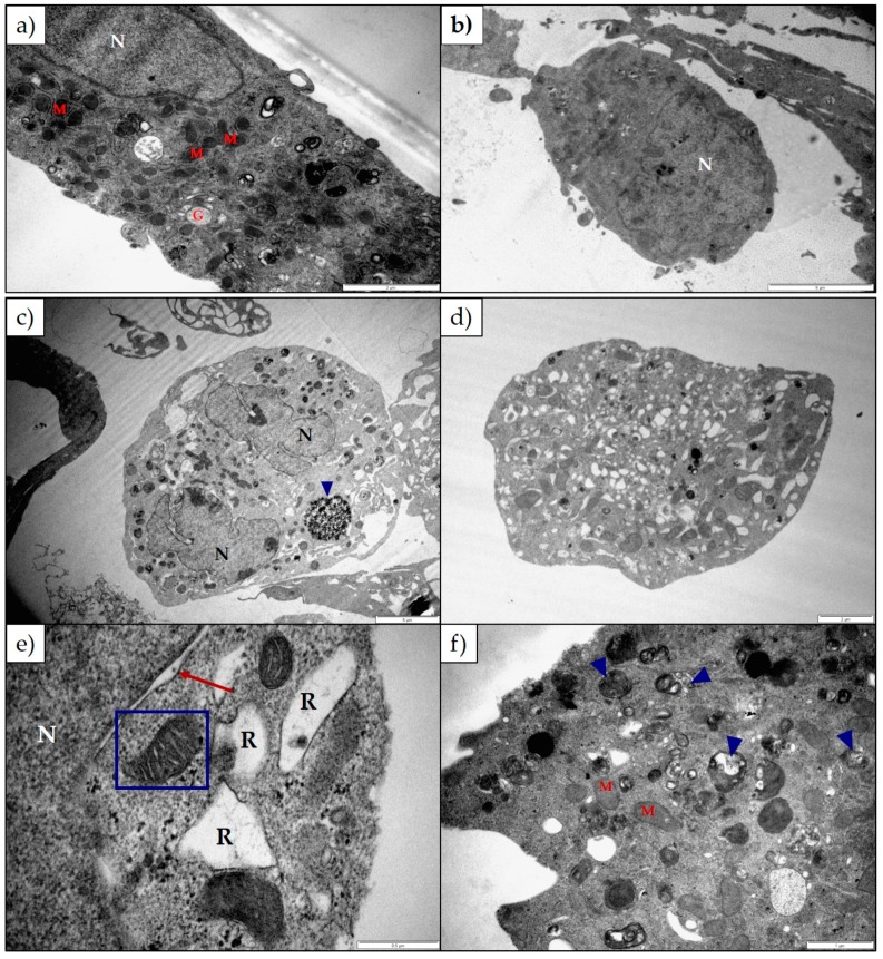
Figure 5.
Electron micrograph of (a,b) untreated Neuro-2a cells and Neuro-2a cells exposed to (c) 100 µM 1α,2α-epoxyscillirosidine for 24 h; (d,e) 100 µM 1α,2α-epoxyscillirosidine for 48 h; (f) 100 µM 1α,2α-epoxyscillirosidine for 72 h. The organelles of untreated Neuro-2a cells appeared normal, with round or slightly convoluted nuclei. Cells exposed to 1α,2α-epoxyscillirosidine were extensively vacuolated, with swollen RER and perinuclear spaces. The cristae of some mitochondria were ballooned, and the nuclei were radially segmented. Many autophagic vesicles could be seen within the cytoplasm (blue arrow heads). G—Golgi complexes; M—Mitochondria; N—Nuclei; R—Rough Endoplasmic Reticulum. The scale bar at the bottom right corner represents 5 µm (b,c), 2 µm (a,d), 1 µm (f) and 0.5 µm (e) respectively.

