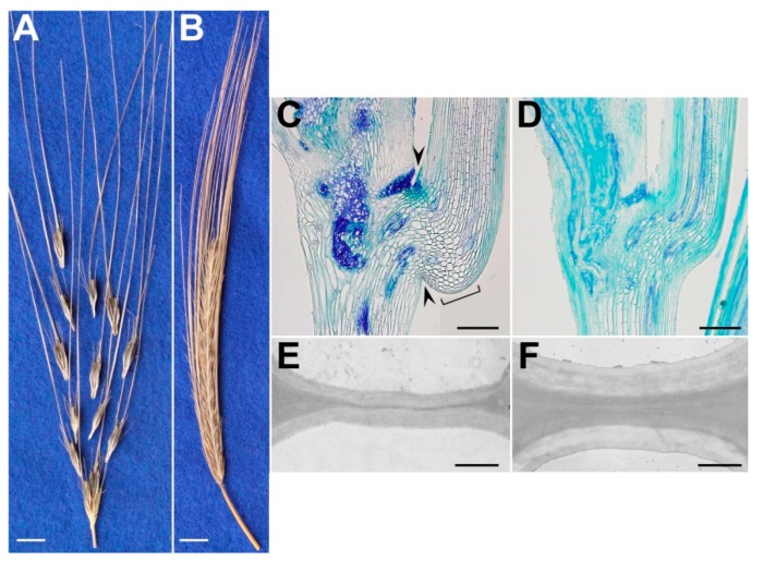
Figure 3.
(A,B) Representative mature spikes of wild barley accession OUH602 (A; brittle) and induced non-brittle rachis mutant M96-1 (B). (C,D) Representative longitudinal sections of junction between two rachis nodes at the anthesis stage, stained with toluidine blue O. Arrowheads: separation layer (or ‘constriction groove’); square bracket: layer of expanded cells. (E,F) Representative transmission electron microscopy showing cell-wall thickness in separation layer of wild (E) and shattering-resistant mutant (F) spikes prior to disarticulation. Scale bars: 1 cm (A,B); 250 µm, (C,D); 1 µm, (E,F) (reproduced with permission from Pourkheirandish et al. [14]; with modifications).

