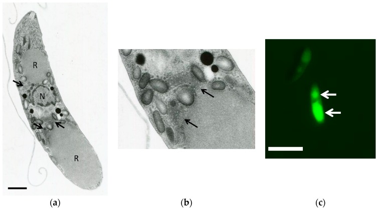
Figure 1.
(a) Electron microscopy of Eimeria tenella sporozoite; (b) high magnification of the mitochondria of the parasite; (c) staining of sporozoites by mitochondrial specific probes (MitoTracker®, Thermo Fisher Scientific). Bars = 2 μm (a) and 5 μm (c). The positions of the mitochondria are indicated by the arrows. R: refractile bodies. N: nucleous.

