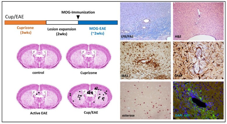
Figure 3.
The Cup/EAE-model (Cuprizone/Experimental autoimmune encephalomyelitis): On the left upper part, the principal experimental setup of a classical Cup/EAE experiment is illustrated. During the first three weeks, animals were intoxicated with cuprizone (0.25%; orange bar), followed by two weeks on normal chow (white bar). At the beginning of week six (arrowhead) animals were immunized with MOG35–55 peptide + CFA/PTX (complete freund’s adjuvant/pertusistoxin). The lower images demonstrate the number and local distribution of perivascular infiltrations. On the right site, histopathological characteristics of such perivascular infiltrations are demonstrated. Luxol fast blue (LFB)/periodic acid-Schiff (PAS); Anti-ionized calcium-binding molecule 1 (IBA1); Anti-glial fibrillary acidic protein (GFAP); Anti-amyloid beta (A4) precursor protein (APP).

