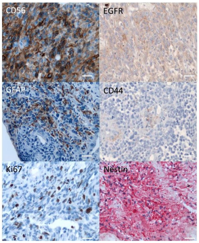
Figure 1.
Original tumor was labelled with different markers (see Table 1) as described in the Materials and Methods, using a revelation DAB (peroxidase) for CD56, CD44, GFAP, EGFR, and Ki67 and a revelation Fast Red (alcaline phosphatase) for Nestin. Only positive labellings are shown. Pictures were captured using Leica ICC50 camera connected to a Leica DM2500 microscope (objective ×20). Scale bar: 100 µm.

