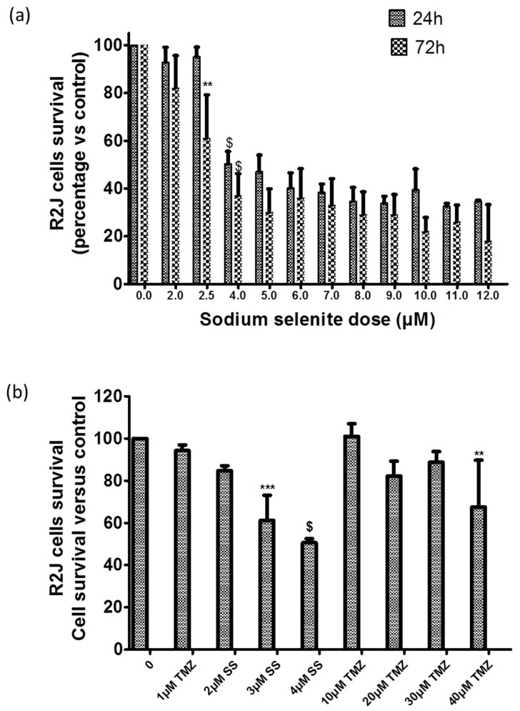
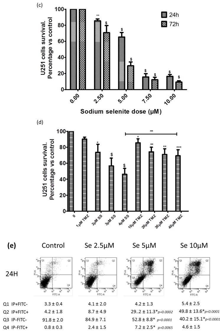
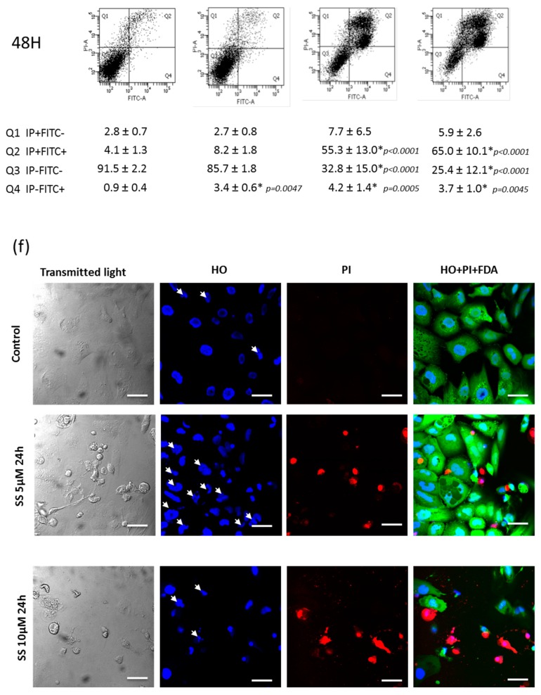
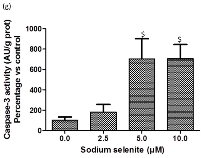
Figure 5.
Sodium selenite (SS) cytotoxicity and cell death triggered. SS is more cytotoxic than TMZ in R2J ((a) and (b)) and in U251 ((c) and (d)) cells: Cell survival was evaluated by an MTT assay performed on cell growing without (control = 100%) or with variable doses of sodium selenite for 24 h and 72 h (a,c) or sodium selenite or TMZ for 72 h, followed by 72 h of wash-out (b,d). Results, expressed in percentage of cell survival vs. the control, are the mean ± SD of three independent experiments with * p < 0.05, ** p < 0.005, *** p < 0.0005, and $ p < 0.0001 vs. control. In (a), only the first statistical significance was specified to avoid an overloading of the graph. (e) Apoptosis (Annexin-V labelling) and necrosis (PI labelling) were evaluated by flow cytometry analysis, 24 h and 48 h after SS treatment. Results are the mean ± SD of four independent experiments. The percentage of cells in each quadrant was compared as a function of the SS concentration and when significant, the p value was informed. Q3 represents viable cells, Q1: Necrotic cells, Q2: Both necrotic and apoptotic cells, and Q4: Apoptotic cells. (f) Confocal microscopy: R2J were exposed to different concentrations of sodium selenite for 24 h and labelling was compared to the controls. Intact cells were revealed by intracellular green fluorescence of FDA (no loss of plasma membrane integrity) whereas the nuclei of necrotic cells were labelled with propidium iodide (PI) (red fluorescence). The Hoechst blue fluorescence allowed the discrimination of apoptotic nuclei with an irregular shape (informed white arrows) that exhibited bean-like morphology or were fragmented whereas the intact nuclei displayed a regular shape. Pictures are representative of three independent experiments. Scale Bar: 40 µm. (g) SS triggered apoptosis via caspase-3. Caspase 3 assay was performed in R2J cells seeded at 6000 cells/cm² and treated with SS for 24 h. Cells and medium were then recovered, centrifuged for 3 min, 360 g, room temperature and rinsed twice with PBS. Cells were lysed in 50 µL of the Caspase-3 lysis buffer. Results are mean ± SD of three independent experiments with $ p < 0.0001 versus the control.


