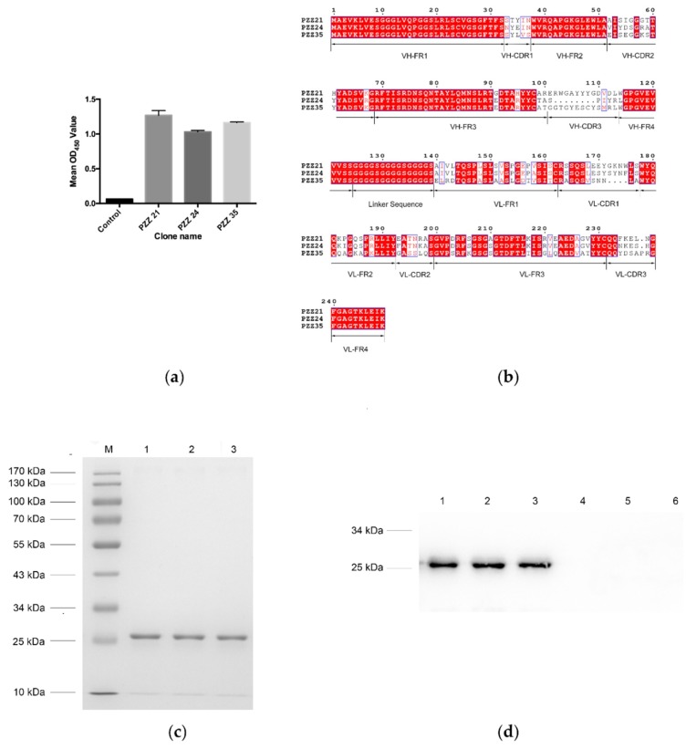
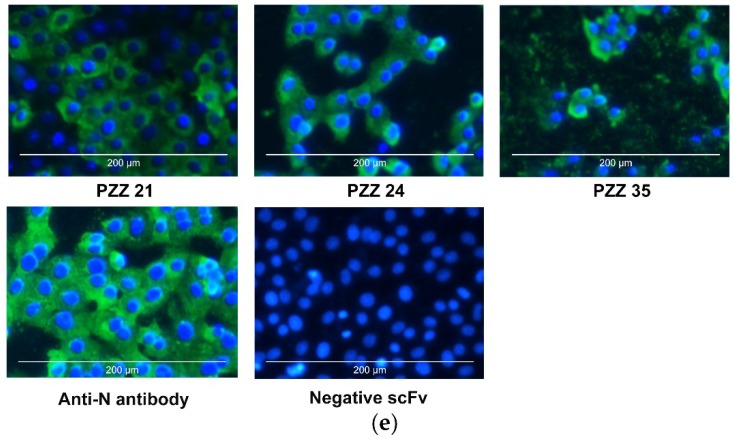
Figure 1.
Identification of scFvs bound to porcine epidemic diarrhea virus (PEDV) antigen. (a) The single chain fragment variable (scFv) obtained from the biopanning process were further analyzed by phage ELISA. An scFv that showed no affinity to PEDV antigen was used as a negative control. Three scFvs with relatively higher values were subjected to further analysis. These scFvs were designated PZZ 21, PZZ 24, and PZZ 35, respectively. (b) The deduced amino acid sequences of PZZ 21, PZZ 24, and PZZ 35 were aligned using ClustalX (version 2.1, www.clustal.org/), and the figure was produced using ESpript (espript.ibcp.fr/). The dots (.) represent the missing amino acids. (c) The PZZ 21, PZZ 24, and PZZ 35 gene sequences were ligated into the expression vector, and the secreted recombinant antibody was purified using Ni-NTA resin and subjected to SDS-PAGE analysis. Lane M: protein molecular weight marker; Lane 1: purified PZZ 21; Lane 2: purified PZZ 24; Lane 3: purified PZZ 35. (d) Purified scFv PZZ 21, PZZ 24, and PZZ 35 were further confirmed by western blot analysis. A mouse anti-His monoclonal antibody was used as the primary antibody. Lane 1: PZZ 21 induced by IPTG; Lane 2: PZZ 24 induced by IPTG; Lane 3: PZZ 35 induced by IPTG; Lane 4: PZZ 21 not induced by IPTG; Lane 1: PZZ 24 not induced by IPTG; Lane 6: PZZ 35 not induced by IPTG. (e) PZZ 21, PZZ 24, and PZZ 35 bound to PEDV-infected cells. PEDV-infected Vero E6 cells were incubated with PZZ 21, PZZ 24, or PZZ 35 at 24 h post-infection and then stained with the FITC conjugated anti-His monoclonal antibody. PEDV-infected cells stained with anti-N PEDV polyclonal antibody were used as the positive control. Stained cells were observed under a fluorescence microscope.


