Figure 3.

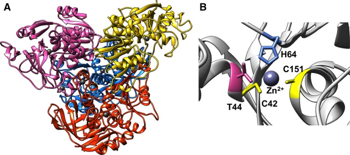
Structure of the homology model of ADH/A1a. (A) Tetrameric structure of ADH/A1a. Monomers are indicated by different colors; Zn ions are dark gray. (B) Conserved catalytic Zn‐binding motif cys42‐his64‐cys151 of ADH/A1a.

Structure of the homology model of ADH/A1a. (A) Tetrameric structure of ADH/A1a. Monomers are indicated by different colors; Zn ions are dark gray. (B) Conserved catalytic Zn‐binding motif cys42‐his64‐cys151 of ADH/A1a.