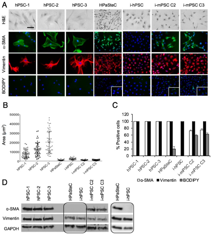
Figure 1.
Phenotypic characterization of pancreatic stellate cells. (A) For morphological analysis, cells were stained with hematoxylin and eosin (H&E), BODIPY for detection of cytoplasmic lipid droplets and immunostained with anti-α-SMA (green) and anti-vimentin (red) antibodies. Nuclei were stained with DAPI (blue). Scale bar = 100 µM. (B) Cell size of the various PSC cultures was determined by measurement of the area of 50 cells for each PSC culture using FIJI software. (C) Number of positive cells for α-SMA, vimentin, and BODIPY in percentage. (D) Cells were lysed and proteins subjected to immunoblotting using anti-α-SMA and anti-vimentin antibodies. Due to high exposure time required for detection, α-SMA expression in HPaSteC cells is also presented in a separate blot. GAPDH was used as a loading control. PSC, pancreatic stellate cell; hPSC, human primary PDAC-derived PSC culture; HPaSteC, PSCs from normal human pancreas; i-hPSC, immortalized human PSCs; i-mPSC C2 and C3, immortalized mouse PSCs clone 2 and 3.

