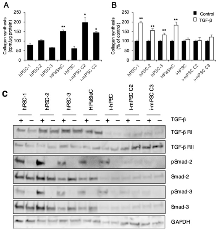
Figure 3.
Collagen synthesis. (A) Basal collagen synthesis. (B) Collagen synthesis after TGF-β (10 nM) stimulation for 24 h. Measurement based on incorporation of [3H]-proline into collagen. Data are mean ± SEM of triplicate determinations. (C) Cells treated with or without TGF-β for 24 h were lysed and proteins subjected to immunoblotting using antibodies against TGF-β receptor I and II, phospho-Smad-2, Smad-2, phospho-Smad-3, and Smad-3. GAPDH was used as loading control. * p < 0.05, ** p < 0.01 comparing average of hPSCs with HPaSteC, i-mPSCs for (A); ** p < 0.01 comparing control (non-treated) cells with TGF-β treated cells for (B). PSC, pancreatic stellate cell; hPSC, human primary PDAC-derived PSC culture; HPaSteC, PSCs from normal human pancreas; i-hPSC, immortalized human PSCs; i-mPSC C2 and C3, immortalized mouse PSCs clone 2 and 3.

