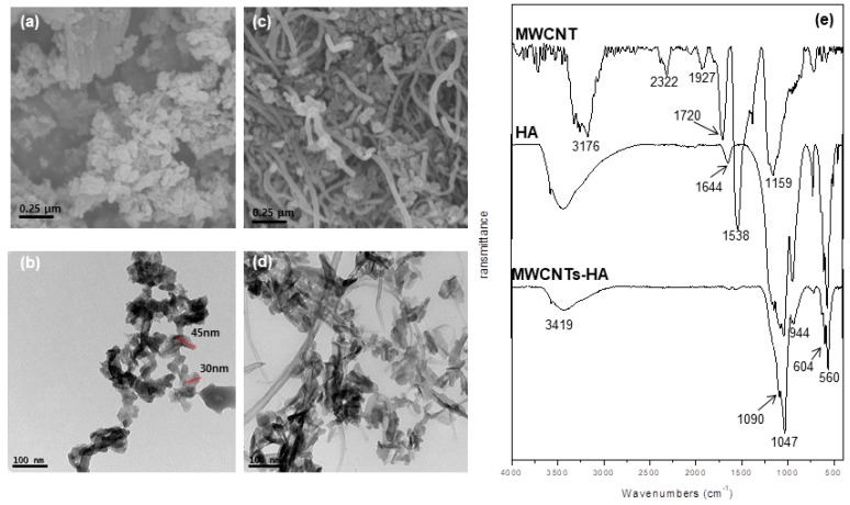
Figure 1.
(a) Field emission scanning electron microscope (FE-SEM) and (b) transmission electron microscopy (TEM) image of sol-gel synthesized hydroxyapatite (HA) powders, (c) FE-SEM image and (d) TEM image of multi walled carbon nanotubes-hydroxyapatite (MWCNTs-HA) powders, and (e) Fourier-transform infrared spectroscopy (FT-IR) spectra of MWCNT, HA and MWCNTs-HA powders.

