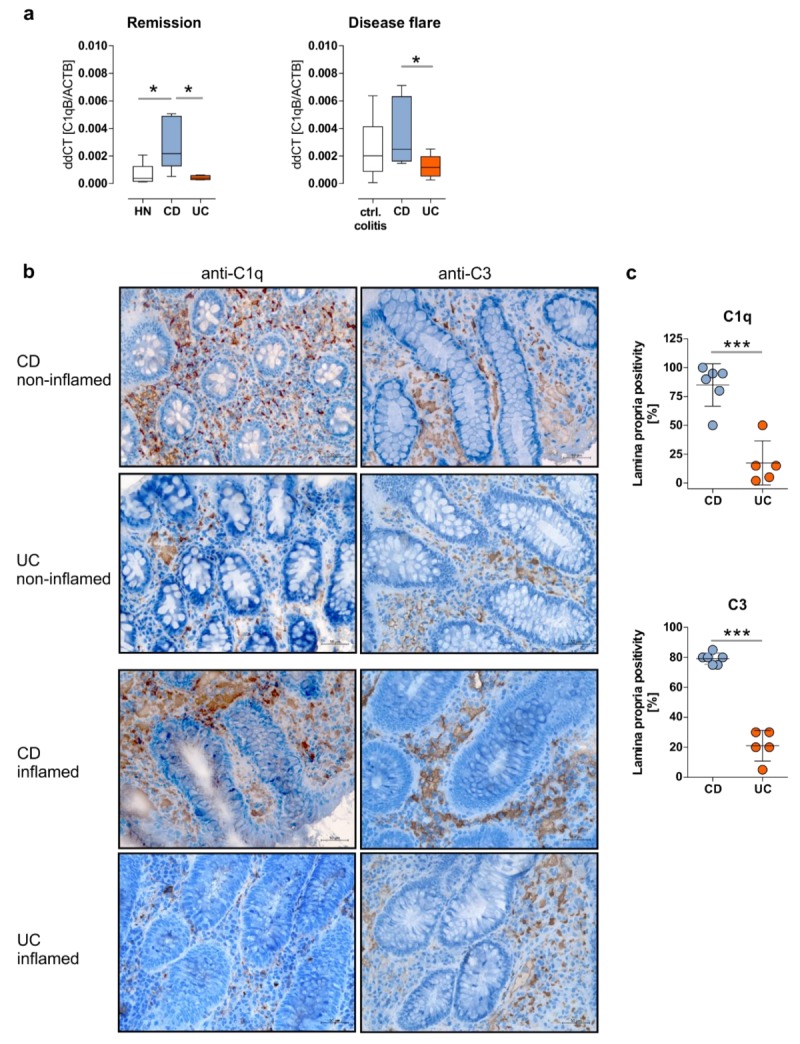
Figure 2.

CD patients in remission display an over-representation of mucosal C1q and C3 expression. (a) The mRNA expression of C1QB in non-inflamed (ni) sigmoidal biopsy samples from IBD patients in remission or HNs, as well as from inflamed sigmoidal biopsy samples from IBD patient with active disease or control colitis patients was analyzed by qPCR using C1QB specific oligonucleotides. ddCt values are depicted. * p ≤ 0.05. (b) Representative pictures of C1q (left panels) or C3 staining (right panels) in colonic tissues from CD or UC patients (non-inflamed or inflamed) (original magnification: 20×). (c) Percentages of C1q or C3 positivity of the lamina propria were determined in analyzed CD (n = 6) or UC (n = 5) tissue samples, irrespectively of the inflammatory status. *** p ≤ 0.001.
